Author Affiliations
Abstract
1 School of Astronautics, Harbin Institute of Technology, Harbin 150000, P. R. China
2 School of Information Science and Engineering, Harbin Institute of Technology, Weihai 264200, P. R. China
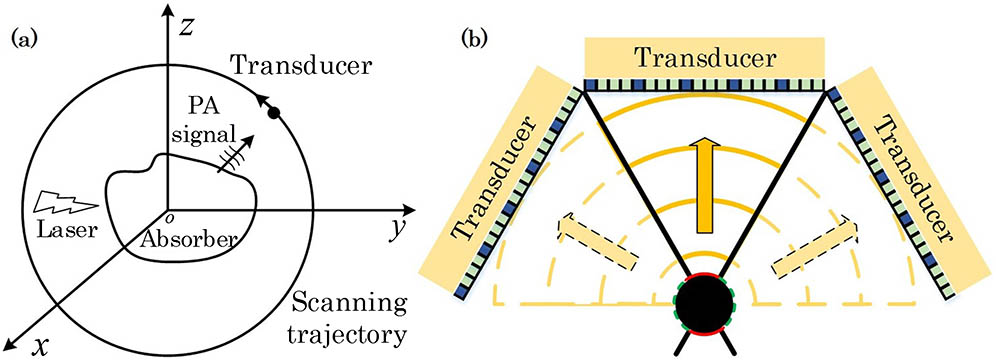
Elastography can be used as a diagnostic method for quantitative characterization of tissue hardness information and thus, differential changes in pathophysiological states of tissues. In this study, we propose a new method for shear wave elastography (SWE) based on laser-excited shear wave, called photoacoustic shear wave elastography (PASWE), which combines photoacoustic (PA) technology with ultrafast ultrasound imaging. By using a focused laser to excite shear waves and ultrafast ultrasonic imaging for detection, high-frequency excitation of shear waves and noncontact elastic imaging can be realized. The laser can stimulate the tissue with the light absorption characteristic to produce the thermal expansion, thus producing the shear wave. The frequency of shear wave induced by laser is higher and the frequency band is wider. By tracking the propagation of shear wave, Young’s modulus of tissue is reconstructed in the whole shear wave propagation region to further evaluate the elastic information of tissue. The feasibility of the method is verified by experiments. Compared with the experimental results of supersonic shear imaging (SSI), it is proved that the method can be used for quantitative elastic imaging of the phantoms. In addition, compared with the SSI method, this method can realize the noncontact excitation of the shear wave, and the frequency of the shear wave excited by the laser is higher than that of the acoustic radiation force (ARF), so the spatial resolution is higher. Compared to the traditional PA elastic imaging method, this method can obtain a larger imaging depth under the premise of ensuring the imaging resolution, and it has potential application value in the clinical diagnosis of diseases requiring noncontact quantitative elasticity.
Elastography shear wave photoacoustic Young’s modulus Journal of Innovative Optical Health Sciences
2024, 17(3): 2350031
Author Affiliations
Abstract
School of Astronautics, Harbin Institute of Technology, Harbin, Heilongjiang 150000, P. R. China
Photoacoustic imaging (PAI) is a noninvasive emerging imaging method based on the photoacoustic effect, which provides necessary assistance for medical diagnosis. It has the characteristics of large imaging depth and high contrast. However, limited by the equipment cost and reconstruction time requirements, the existing PAI systems distributed with annular array transducers are difficult to take into account both the image quality and the imaging speed. In this paper, a triple-path feature transform network (TFT-Net) for ring-array photoacoustic tomography is proposed to enhance the imaging quality from limited-view and sparse measurement data. Specifically, the network combines the raw photoacoustic pressure signals and conventional linear reconstruction images as input data, and takes the photoacoustic physical model as a prior information to guide the reconstruction process. In addition, to enhance the ability of extracting signal features, the residual block and squeeze and excitation block are introduced into the TFT-Net. For further efficient reconstruction, the final output of photoacoustic signals uses ‘filter-then-upsample’ operation with a pixel-shuffle multiplexer and a max out module. Experiment results on simulated and in-vivo data demonstrate that the constructed TFT-Net can restore the target boundary clearly, reduce background noise, and realize fast and high-quality photoacoustic image reconstruction of limited view with sparse sampling.
Deep learning feature transformation image reconstruction limited-view measurement photoacoustic tomography Journal of Innovative Optical Health Sciences
2024, 17(3): 2350028
Author Affiliations
Abstract
1 School of Astronautics, Harbin Institute of Technology, Harbin 150000, P. R. China
2 Weihai Institute of Product Quality Standards and Metrology, Weihai 264200, P. R. China
Photoacoustic imaging (PAI) has been developed, and photoacoustic computed tomography (PACT) is widely used for in vivo tissue and mouse imaging. Simulated annealing (SA) algorithm solves optimization problems, and compressed sensing (CS) recovers sparse signals from undersampled measurements. We aim to develop an advanced sparse imaging framework for PACT, which invloves the use of SA to find an optimal sparse array element distribution and CS to perform sparse imaging. PACT reconstructions were performed using a dummy and porcine liver phantoms. Compared to traditional sparse reconstruction algorithms, the proposed method recovers signals using few ultrasonic transducer elements, enabling high-speed, low-cost PACT for practical application.Photoacoustic imaging (PAI) has been developed, and photoacoustic computed tomography (PACT) is widely used for in vivo tissue and mouse imaging. Simulated annealing (SA) algorithm solves optimization problems, and compressed sensing (CS) recovers sparse signals from undersampled measurements. We aim to develop an advanced sparse imaging framework for PACT, which invloves the use of SA to find an optimal sparse array element distribution and CS to perform sparse imaging. PACT reconstructions were performed using a dummy and porcine liver phantoms. Compared to traditional sparse reconstruction algorithms, the proposed method recovers signals using few ultrasonic transducer elements, enabling high-speed, low-cost PACT for practical application.
Photoacoustic computed tomography sparse simulated annealing compressed sensing Journal of Innovative Optical Health Sciences
2022, 15(5): 2250030
1 哈尔滨工业大学(威海)信息科学与工程学院,山东 威海 264209
2 哈尔滨工业大学航天学院,黑龙江 哈尔滨 150001
3 威高集团有限公司,山东 威海 213000
4 中国科学院苏州生物医学工程技术研究所,江苏 苏州 215163
光声层析成像是一种非侵入式的医学成像技术,与其他成像方法相比具备诸多优势,可以为肿瘤早期诊断提供新的成像思路。对光声信号的分析与去噪能提高成像系统的信噪比(SNR)和成像质量。为此,提出了一种针对光声信号的智能去噪算法。首先,利用自适应白噪声完备集合经验模态分解完成光声信号的分解;其次,采用小波阈值去噪方法完成对特定模态光声信号的高频去噪;最后,利用K奇异值分解对预处理后的光声信号进行稀疏重构,实现光声信号的智能去噪。仿真和实验结果表明,所提算法在SNR和均方根误差(RMSE)等方面相比于其他去噪算法均有改善,可以有效去除三维肿瘤仿体光声重建图像中的噪点与伪影,并保留图像的边缘信息。所提智能去噪算法能根据含噪光声信号的特征自适应地去噪,达到更好的去噪效果,可以作为一种成像前的辅助手段应用于光声成像领域。
成像系统 光声层析成像技术 经验模态分解 小波阈值 K奇异值分解 噪声 激光与光电子学进展
2022, 59(8): 0811006
1 哈尔滨工业大学(威海)信息科学与工程学院, 山东 威海 264209
2 哈尔滨工业大学航天学院, 黑龙江 哈尔滨 150000
光热治疗是一种非侵入式、靶向性的新型治疗技术,但现有的光热治疗技术不能实时监测靶区的温度分布,且开环的激光控制方式不仅增大了治疗难度,也会对病灶周边的正常组织造成不可逆损伤。为此,提出了一种基于光声温度精准调控的光热治疗方法。研究了基于光声图像的温度成像算法,提出了光声温度敏感因子的概念,设计了基于光声温度敏感因子的闭环温度控制算法,最后搭建了一套基于光声温度精准调控的新型光热治疗系统,并进行了仿体实验。实验结果表明:基于光声温度精准调控的光热治疗方法可实现靶区温度的非接触式精准测量与控制,系统调节时间在10 s以内且温度控制均方根误差在0.7 ℃以内。基于光声温度精准调控的光热治疗方法可以作为一种更精准、高效的辅助手段应用于光热治疗领域。
医用光学 光热治疗 光声测温 敏感因子 精准调控 中国激光
2020, 47(10): 1007001
1 哈尔滨工业大学(威海)信息科学与工程学院, 山东 威海 264209
2 中国科学院深圳先进技术研究院生物医学光学与分子影像研究室, 广东 深圳 518055
3 哈尔滨工业大学航天学院, 黑龙江 哈尔滨 150001
消化道肿瘤是最常见的肿瘤疾病之一。新兴的光声成像技术可以敏锐地捕捉肿瘤周围滋养血管的信息,有助于临床进行更精准的诊断。匹配的血管增强算法可以有效突出图像中的血管网络,但光声活体内窥成像的探测角度有限,易造成明显的血管形态异常,现有方法很难实现有效的血管增强。采用自研的光声内窥系统对大鼠直肠进行活体成像,针对活体成像结果提出了一种融合结构和强度两个层面信息的三维血管增强算法,并采用该算法对结直肠血管图像进行了增强。结果表明:所提算法可以有效提升增强效果,抑制机械抖动带来的边缘毛刺,在活体状态下获取了高质量的结直肠三维血管图像,说明其在基础医学研究和临床应用中具有一定的潜在价值。
医用光学 内窥成像 光声成像 血管增强 三维增强
Author Affiliations
Abstract
1 Department of Control Science and Engineering, Harbin Institute of Technology, Harbin Heilongjiang 150001, P. R. China
2 Department of Bioengineering, University of Pittsburgh, Pittsburgh, PA 15213, USA
The synthetic aperture-based linear-array photoacoustic tomography (PAT) was proposed to address the limited-view shortcomings of the single aperture, but the detection field of view (FOV) determined by the aperture orientation effect was not fully considered yet, leading to the limited-view observation and image resolution degradation. Herein, the aperture orientation effect was proposed from the theoretical model and then it was verified via both the numerical simulation and phantom experiment. Different orientations were enumerated sequentially in the simulation to approximate the ideal full-view case for the optimal detection FOV, considering the detection pattern of the linear-array transducer. As a result, the corresponding optimal aperture orientation was 60� if the synthetic aperture was seamlessly established by three single linear arrays, where the overlapped detection pattern was optimized from the individual linear-array transducer at the adjacent positions. Therefore, the limited-view artifacts were minimized and the image resolution was enhanced in this aperture orientation. This study showed that the aperture orientation had great influence on the optimal detection FOV in the synthetic aperture configuration, where the full-view imaging quality and enhanced image resolution could be achieved.
Photoacoustic tomography synthetic aperture aperture orientation linear-array transducer Journal of Innovative Optical Health Sciences
2018, 11(4): 1850015
Author Affiliations
Abstract
1 Department of Control Science and Engineering, Harbin Institute of Technology, Harbin 150001, China
2 Department of Gastroenterology, General Hospital of Chinese People’s Armed Police Forces, Beijing 100039, China
3 Institute of Opto-electronics, Harbin Institute of Technology, Harbin 150080, China
Photoacoustic tomography (PAT) has the unique capability of visualizing optical absorption inside several centimeters-deep biological tissue with a high spatial resolution. However, single linear-array transducer-based PAT suffers from the limited-view challenge, and thus the synthetic aperture configuration is designed that still requires multichannel data acquisition hardware. Herein, a feasible synthetic aperture PAT based on compressed sensing reconstruction is proposed. Both the simulation and experimental results tested the theoretical model and validated that this approach can improve the image resolution and address the limited-view problem while preserving the target information with a fewer number of measurements.
110.5120 Photoacoutic imaging 100.3020 Image reconstruction-restoration 170.5120 Photoacoustic imaging Chinese Optics Letters
2017, 15(10): 101102
Author Affiliations
Abstract
Department of Control Science and Engineering, Harbin Institute of Technology, Harbin 150001, China
Photoacoustic tomography is a noninvasive and nonionized biomedical imaging modality but it cannot reveal the inner structure and sideward boundary information of blood vessels in the linear array detection mode. In contrast, Monte Carlo (MC) light transport could provide the optical fluence distribution around the entire vascular area. This research explores the combination of linear array transducer-based photoacoustic tomography and MC light transport in the blood vessel quantification. Simulation, phantom, and in vivo experiments are in good correlation with the ultrasound imaging, validating this approach can clearly visualize the internal region of blood vessels from background tissue.
170.5120 Photoacoustic imaging 100.3020 Image reconstruction-restoration 110.5120 Photoacoutic imaging Chinese Optics Letters
2017, 15(11): 111701
Author Affiliations
Abstract
Department of Control Science and Engineering, Harbin Institute of Technology, Harbin 150001, China
As a high-resulotion biological imaging technology, photoacoustic microscopy (PAM) is difficult to use in real-time imaging due to the long data acquisition time. Herein, a fast data acquisition and image recovery method named sparse PAM based on a low-rank matrix approximation is proposed. Specifically, the process to recover the final image from incomplete data is formulated into a low-rank matrix completion framework, and the “Go Decomposition” algorithm is utilized to solve the problem. Finally, both simulated and real PAM experiments are conducted to verify the performance of the proposed method and demonstrate clinical potential for many biological diseases.
100.3020 Image reconstruction-restoration 170.5120 Photoacoustic imaging 170.0110 Imaging systems Chinese Optics Letters
2016, 14(9): 091701


